Physio Hub:
Evidence-Based Article
Hip Labral Tears in Athletes
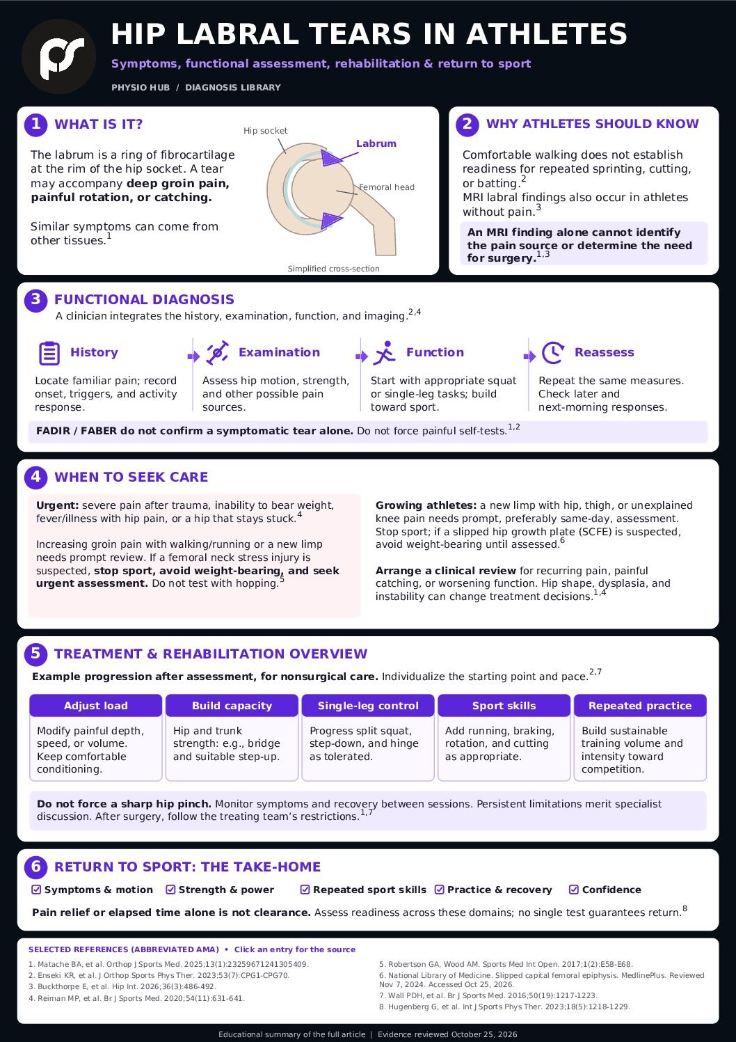
A hip labral tear is an injury to the ring of tissue around the hip socket. It can accompany deep groin pain, painful rotation, catching, or difficulty tolerating sport. The same finding can also appear on MRI in someone without hip pain. An athlete therefore needs an assessment that connects the scan with symptoms, examination findings, and sporting demands. [1,2]
For athletes, coaches, parents, and clinicians, the practical questions are whether the hip joint is contributing to symptoms, which functions are limited, and what will restore dependable participation and performance. Many athletes can begin with rehabilitation. Others benefit from specialist assessment of an associated problem such as impingement or instability.[3,4]
This article explains that decision process. The rehabilitation examples are intended for an assessed, nonoperative presentation; postoperative care requires a procedure-specific plan.
What is a Hip Labral Injury
The role of the labrum
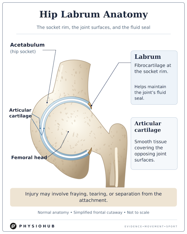
The acetabular labrum is a ring of fibrocartilage attached to the rim of the acetabulum, the socket of the hip. It helps maintain a fluid seal around the femoral head and contributes to normal joint mechanics. It is different from the articular cartilage that covers the joint surfaces. Laboratory research demonstrates the importance of the seal, but a laboratory finding cannot tell us how much pain an individual tear will cause.[5]
A labral injury may involve fraying, tearing, or separation from its attachment. Symptoms may begin after a traumatic twist or emerge gradually, and labral injuries may coexist with other hip conditions. The word “tear” describes tissue appearance; its clinical significance depends on the whole presentation.[4,6]
Associated hip conditions
Several conditions can overlap with labral pathology:
- Femoroacetabular impingement syndrome, or FAIS: symptoms and clinical signs occur alongside relevant hip shape findings. Cam morphology refers to a less rounded junction between the femoral head and neck; pincer morphology describes acetabular overcoverage. Having one of these shapes alone does not establish FAIS.[7]
- Acetabular dysplasia or hip instability: insufficient socket coverage or inadequate joint restraint can change how the hip is loaded. Microinstability is a clinical diagnosis requiring a combination of findings; flexibility alone does not establish it.[8]
- Cartilage injury or degenerative change: these may coexist and influence treatment options and prognosis. A tear should be considered within the condition of the joint as a whole.[4]
Typical symptoms
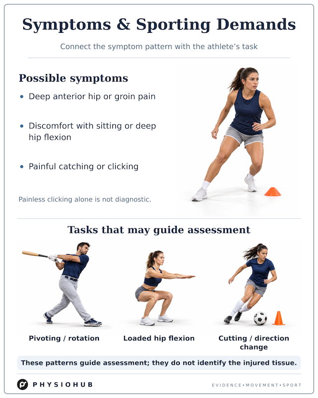
Possible symptoms include deep anterior hip or groin pain, discomfort with prolonged sitting or deep hip flexion, and pain with pivoting or rotation. Some athletes describe painful catching or clicking. These features raise suspicion, but they also occur with other conditions. Painless clicking alone is insufficient to diagnose a symptomatic tear.[6,7]
Why Is It Important in Athletes?
An athlete may tolerate walking yet struggle with repeated acceleration, cutting, landing, or rotation. A baseball hitter’s pivot, a catcher’s squat, and a soccer player’s change of direction are examples of different tasks that can guide assessment. They do not identify the injured tissue by themselves.
An imaging label also requires context. A 2026 study of 166 soccer and Australian football players found no difference between symptomatic and asymptomatic groups in the prevalence of the assessed MRI abnormalities. This does not mean that labral injuries never cause pain; it means that imaging alone cannot reliably explain an individual athlete’s symptoms.[2]
The same issue matters in younger athletes. In a 2025 study, labral tears appeared on MRI in 18% of 50 children and adolescents without hip symptoms and in 30% of 50 with symptoms. The difference was not statistically significant. The asymptomatic group was recruited through attendance for knee MRI, so these figures should not be treated as prevalence estimates for every youth sport population.[9]
Recovery should also be defined carefully. Returning to some sport participation, returning to the previous competitive level, and performing at the previous standard are different outcomes. Athlete research after hip arthroscopy shows why broad “return to sport” statistics can conceal ongoing limitations.[10]
Functional Diagnosis of Hip Labral Tears
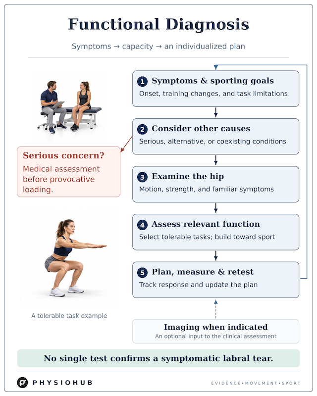
Functional diagnosis means identifying the activities the athlete cannot currently tolerate, measuring relevant impairments, and testing which changes improve performance or symptoms. It complements the medical diagnosis. A squat, strength test, or movement correction cannot independently confirm that a labral tear is the pain source.[3,4]
Step 1 Establish the symptom pattern and sporting goal
Ask where the pain is felt, how it began, and which activities reproduce the athlete’s familiar symptoms. Clarify whether there was a sudden injury or a gradual increase after a change in training. Record the response during activity, later that day, and the following morning.
Distinguish a harmless noise from painful catching and from true locking, where the joint becomes mechanically stuck. Ask about limping, difficulty bearing weight, pain at rest, previous hip problems, and the specific sporting task the athlete wants to recover. The assessment must first consider serious or nonmusculoskeletal causes when the history suggests them.[4]
For a runner with increasing weight-bearing groin pain, training changes and bone-health factors matter. A suspected bone stress injury should be assessed before hopping, running, or other provocative performance testing.[11]
Step 2 Consider competing and coexisting diagnoses
Hip and groin pain can arise from several neighboring structures. Examination may identify adductor-related, iliopsoas-related, inguinal-related, or pubic-related groin pain, and more than one condition may be present.[12]
| Possible contributor | Examples of findings that guide assessment |
| Adductor-related groin pain | Recognizable pain with adductor palpation and resisted adduction |
| Iliopsoas-related pain or snapping | Anterior symptoms associated with hip-flexor loading or reproducible snapping |
| Inguinal-related or pubic-related pain | Local tenderness and symptoms with abdominal or sporting loads |
| Lumbar or other referred pain | Symptoms influenced by spinal examination or accompanied by neurological findings |
| Bone stress injury or a growth-related hip disorder | Increasing weight-bearing pain, a limp, or an adolescent presenting with hip, thigh, or knee pain |
These patterns guide the differential diagnosis; they do not replace a full examination. The first three categories follow the Doha framework, while spinal and serious hip conditions require their own assessment.[4,12]
Step 3 Examine the hip and interpret special tests cautiously
Measure hip motion in a reproducible position and note both the range and the symptom response. A painful restriction is different from a comfortable anatomical limit. Assess strength where relevant, including hip flexion, extension, abduction, adduction, and rotation, alongside trunk and lower-limb function.[3]
Common examination components include:
| Examination component | Useful information | Main limitation |
| FADIR | Whether combined flexion, adduction, and internal rotation reproduce familiar symptoms | A positive result does not identify the labrum as the source |
| FABER | Symptom location and tolerance of flexion, abduction, and external rotation | Pain may arise from different structures; the location matters |
| Hip range of motion | Pain, restriction, asymmetry, and change over time | Restricted rotation alone does not diagnose a tear or FAIS |
| Resisted muscle testing | Pain with loading and measurable force capacity | Weakness may be a contributor, consequence, adaptation, or separate finding |
In a diagnostic study of FAIS, FADIR had relatively high sensitivity but low specificity, illustrating why a positive provocation test needs context. Those results concern FAIS and cannot simply be applied as a diagnostic rule for isolated labral tears.[13]
There is no universally validated athlete-specific cluster that confirms a symptomatic labral tear. Anterior groin pain, painful deep flexion, and catching with rotation can support suspicion, but they are not a stand-alone prediction rule.[1]
Athletes should not repeatedly force these positions at home to “check the labrum.” A clinician can select and interpret the tests while considering other diagnoses.
Step 4 Assess the function that matters
Once higher-risk conditions have been excluded, choose tasks that match the athlete’s current capacity. Examples include a squat, step-down, single-leg squat, supported hinge, or balance task. Later assessment may include running, acceleration, deceleration, jumping, cutting, and sport-specific rotation.[3,14]
For each task, record the symptom location, range, speed, load, movement strategy, and ability to repeat it. A visible asymmetry is an observation to investigate, not proof of the injury mechanism. Changing depth, stance, tempo, or external load can help identify a more tolerable starting point.
For example, a hitter who tolerates a controlled split squat but develops groin pain during fast batting practice has a different functional problem from someone who cannot walk comfortably. The assessment should connect the exercise capacity to the actual task rather than treating completion of an exercise as the endpoint.
Step 5 Use imaging to answer a clinical question
When a clinician suspects hip joint pathology, radiographs are commonly the first imaging step. They help evaluate femoral and acetabular shape, socket coverage, arthritis, and other bony conditions. They do not directly show the labrum.[15]
MRI can assess the labrum, cartilage, bone, and nearby soft tissues. MR arthrography adds contrast into the joint. After nondiagnostic radiographs in a patient with a suspected labral tear, the American College of Radiology considers both noncontrast hip MRI and MR arthrography appropriate options. The choice depends on the clinical question, equipment, local expertise, and prior imaging; every patient does not require an arthrogram.[15]
If it remains unclear whether symptoms arise from inside the joint, a clinician may consider an image-guided local anesthetic injection. Symptom relief can support an intra-articular contribution, but cannot isolate the labrum from every other joint structure or guarantee the outcome of surgery.[16,17]
Step 6 Establish a baseline and retest
Use the same meaningful tasks and measurements over time. Patient-reported tools such as the International Hip Outcome Tool, HAGOS, or the Hip Outcome Score sports subscale can complement strength and performance measures.[3]
A useful clinical record connects the athlete’s main task, the current limiting factor, the proposed contributor, the intervention, and the response. For example, improved tolerance after reducing squat depth helps choose an exercise range; it does not prove that the labrum was the original pain generator.
Other clinically relevant information
Clinical improvement and tissue healing are different questions
Symptoms and function can improve during nonsurgical care. That improvement does not establish that a tear has disappeared on MRI. In a nonsurgical labral-tear case series, functional scores improved over one year, but many participants continued to report pain or activity limitations. This supports monitoring the athlete’s actual outcome rather than assuming that either a better score or a persistent imaging finding tells the whole story.[18]
Treatment expectations should therefore include the activities the athlete can perform, symptom behavior, confidence, and the sustainability of training. Persistent restriction still deserves reassessment even if some measures have improved.
When to seek medical care
Seek urgent medical assessment for major trauma with severe hip pain, inability to bear weight, or fever and feeling unwell with significant hip pain. These presentations can indicate conditions requiring prompt investigation beyond a labral tear.[4]
Increasing groin pain with walking or running, a new limp, or progression toward rest pain warrants prompt assessment for bone stress injury. An early X-ray can be normal; when clinical suspicion remains, MRI may be needed. If a femoral neck stress injury is suspected, stop sport and avoid weight-bearing on the affected leg until urgently assessed; crutches or assistance may be needed. Do not test it with running or hopping.[11,19]
In a growing child or adolescent, a new limp with hip, groin, thigh, or unexplained knee pain needs prompt medical assessment, preferably the same day. Slipped capital femoral epiphysis is an important possibility. Stop sport; if this condition is suspected, avoid weight-bearing until evaluated. Do not assume these symptoms are simply growing pains.[20,21]
If the hip becomes stuck and cannot move normally, seek prompt medical assessment and do not force it through the blocked movement. Arrange a sports medicine or physical therapy assessment when pain keeps recurring, painful catching limits activity, normal function is deteriorating, or initial load modification is not producing clear improvement. Persistent symptoms do not automatically mean that surgery is required.
What athletes can monitor themselves
For mild, stable symptoms without the features above, temporarily reduce the specific loads that provoke symptoms while maintaining comfortable activity. Useful adjustments may include a shallower squat, shorter sessions, less repeated pivoting, or a change in cycling position. Record what was done and how the hip responded afterward.[22]
As a practical monitoring guide, reduce the next session’s load if symptoms clearly escalate, a limp appears, or the hip remains substantially worse the following morning. This is a load-adjustment strategy, not a validated test of labral healing. A comfortable session also does not establish readiness for unrestricted competition.
Mobility work should remain tolerable. Repeatedly forcing a sharp anterior pinch or a firm painful end range is not a useful way to restore motion. The Personalised Hip Therapy protocol specifically avoided painful hard-end stretching.[22]
Treatment and Rehabilitation Overview
Begin with an individualized nonoperative plan when appropriate
Education, adjustment of provocative activity, and progressive exercise form a reasonable starting point for many assessed presentations. A supervised trial commonly extends over several months, with regular reassessment. The duration should respond to progress, symptoms, structural considerations, and the athlete’s goals; a fixed waiting period should not delay review of a worsening or atypical presentation.[3,14]
A prospective adolescent FAIS study reported sustained improvements at five years after a staged care pathway beginning with activity modification and physical therapy. Only 51 of the original 100 patients were available at that follow-up, and the pathway also allowed injections and surgery. It should not be interpreted as proof that rehabilitation alone succeeds for every adolescent labral tear.[23]
What recent exercise research adds
The 2026 PhysioFIRST trial compared two physiotherapist-led approaches in 154 adults with FAIS. Targeted strengthening did not outperform standardized stretching on the primary hip-related quality-of-life or global pain and function ratings at six months. A secondary analysis classifying participants as improved or not improved favored strengthening for perceived pain, and strength gains were also greater. Both groups improved in hip-related quality of life. The study compared two active treatments and did not assess structural healing of labral tears.[24]
The practical implication is to select exercises for the athlete’s limitations and sporting demands, then assess the response. Current evidence does not establish one exercise sequence as a universal treatment for all labral injuries.
A practical rehabilitation progression
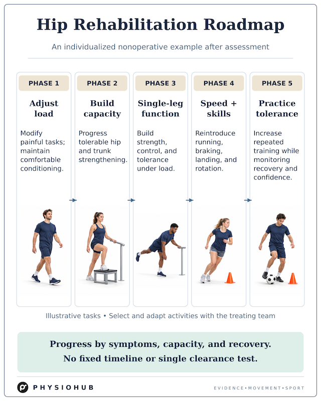
The following is an illustrative progression informed by rehabilitation protocols and clinical reasoning. It is not a validated universal labral-tear program. Select the entry point and progression according to the assessment; sets, repetitions, resistance, and frequency require individual adjustment.[14,22]
Phase 1 Adjust the current load. Identify the painful tasks and temporarily modify their depth, duration, speed, or frequency. Maintain an acceptable form of conditioning. Walking, pool exercise, or cycling may suit some athletes if the position and workload are comfortable.
Phase 2 Rebuild basic capacity. Choose tolerable hip and trunk exercises. Examples include a bridge, suitable hip abduction or adduction work, trunk control exercises, and a controlled step-up. Progress resistance and range when the response supports it; the exercise name itself does not determine suitability.
Phase 3 Restore loaded single-leg function. Examples include a modified squat or split squat, step-down, supported single-leg hinge, balance task, and resisted trunk rotation. Address measurable deficits while allowing a movement strategy that suits the athlete’s anatomy and task.
Phase 4 Reintroduce speed and sport skills. Progress the relevant demands, such as running, acceleration, braking, landing, and rotation. Cutting sports may advance from planned direction changes to reactive tasks. Batting or throwing practice can build from controlled drills toward full-speed repetitions.
Phase 5 Build repeated practice tolerance. Increase exposure toward normal training, including the volume, intensity, and fatigue relevant to the sport. Progression requires a sustainable response across sessions, alongside confidence and performance.
These examples translate broad rehabilitation principles into practical options. They should be modified when the suspected diagnosis, symptoms, or response changes. Manual therapy or other symptom-relief approaches may help selected patients participate in exercise, but do not demonstrate that the labrum has healed.[14]
When surgery enters the discussion
A hip specialist may be helpful when meaningful pain, mechanical symptoms, or sporting limitations persist despite an adequately delivered rehabilitation trial. Earlier review may be appropriate when instability, dysplasia, or another structural problem changes the treatment options.
Two major randomized trials in selected patients with symptomatic FAIS found greater average improvement after arthroscopy than after physiotherapy-based care, although both approaches improved outcomes. The operations often addressed bony impingement and associated labral or cartilage problems. These were not trials showing that every isolated MRI-detected tear should be repaired.[25,26]
The surgical discussion may include repair of suitable labral tissue, reconstruction when tissue cannot be repaired, and treatment of relevant bony or capsular pathology. Dysplasia requires assessment of socket coverage and stability; treating the labrum alone may not address the underlying problem.[1,8]
Potential benefits must be weighed against rehabilitation time and risks such as persistent symptoms, infection, nerve-related symptoms, or further surgery. A randomized trial’s medium-term follow-up did not establish that arthroscopy prevents osteoarthritis: its radiographic joint-space outcome did not differ between groups, and follow-up imaging was incomplete.[25,27]
Injections and other adjuncts
A corticosteroid injection may offer symptom relief in selected circumstances, while local anesthetic may assist diagnostic reasoning. Neither response establishes labral healing. The clinician should review suitability and risks. Evidence for PRP or other orthobiologics is insufficient to promise reliable regeneration of the labrum.[1]
Return to sport and return to performance
Recovery after surgery is usually measured in months. The procedure, healing requirements, sport, and residual deficits affect the timeline. Postoperative weight-bearing and motion restrictions must come from the treating team; the nonoperative examples above should not override them.[14,28]
For either pathway, a return-to-sport discussion should address the following domains:
| Domain | Practical question |
| Symptoms | Are daily activities and progressive training tolerated without a recurring substantial flare? |
| Motion | Is there adequate comfortable motion for the required sporting task? |
| Strength and power | Are measured capacities appropriate for the athlete’s baseline and sport demands? |
| Functional performance | Can the athlete control and repeat relevant running, landing, cutting, or rotational tasks? |
| Training exposure | Can increasing portions of practice be completed with an acceptable recovery response? |
| Confidence and performance | Does the athlete trust the hip, and is performance moving toward the required level? |
These domains reflect proposed clinical frameworks, not a universally validated clearance test. Side-to-side comparisons can be useful, but the other hip may also be weak or symptomatic. Passing a single symmetry threshold does not guarantee safe or successful return.[28]
Build the pathway from modified practice toward full training and competition, then continue to monitor actual performance. Returning to the field is one milestone; restoring the athlete’s previous competitive standard may take longer.[10]
< Summary >
Hip labral injuries require a diagnosis that connects symptoms, examination findings, imaging, and the condition of the whole joint. An MRI finding alone does not tell us whether the athlete needs treatment or which treatment is appropriate.
Functional assessment identifies the tasks that matter and the capacities that need rebuilding. Rehabilitation can help many athletes, while persistent limitations or important structural findings justify specialist discussion. The evidence supporting surgery is strongest for selected symptomatic FAIS populations and should not be generalized to every tear.
For athletes and parents, the next step is to recognize symptoms that warrant assessment, adjust loads sensibly, and track meaningful changes. For clinicians and coaches, the goal is a shared plan that restores sustainable training and performance, with progression guided by the athlete’s response.
<PDF file>
< Reference >
- Matache BA, Belzile EL, Ayeni OR, et al. Management of labral tears in the hip: a consensus statement. Orthop J Sports Med. 2025;13(1):23259671241305409. doi:10.1177/23259671241305409
- Buckthorpe E, Heerey JJ, Entwistle T, et al. What is the prevalence of extra-articular and intra-articular magnetic resonance imaging findings in football players with and without hip and/or groin pain? A cross-sectional study of 166 football players. Hip Int. 2026;36(3):486-492. doi:10.1177/11207000261416847
- Enseki KR, Bloom NJ, Harris-Hayes M, et al. Hip pain and movement dysfunction associated with nonarthritic hip joint pain: a revision. J Orthop Sports Phys Ther. 2023;53(7):CPG1-CPG70. doi:10.2519/jospt.2023.0302
- Reiman MP, Agricola R, Kemp JL, et al. Consensus recommendations on the classification, definition and diagnostic criteria of hip-related pain in young and middle-aged active adults from the International Hip-related Pain Research Network, Zurich 2018. Br J Sports Med. 2020;54(11):631-641. doi:10.1136/bjsports-2019-101453
- Philippon MJ, Nepple JJ, Campbell KJ, et al. The hip fluid seal—Part I: the effect of an acetabular labral tear, repair, resection, and reconstruction on hip fluid pressurization. Knee Surg Sports Traumatol Arthrosc. 2014;22(4):722-729. doi:10.1007/s00167-014-2874-z
- Burnett RS, Della Rocca GJ, Prather H, Curry M, Maloney WJ, Clohisy JC. Clinical presentation of patients with tears of the acetabular labrum. J Bone Joint Surg Am. 2006;88(7):1448-1457. doi:10.2106/JBJS.D.02806
- Griffin DR, Dickenson EJ, O’Donnell J, et al. The Warwick Agreement on femoroacetabular impingement syndrome (FAI syndrome): an international consensus statement. Br J Sports Med. 2016;50(19):1169-1176. doi:10.1136/bjsports-2016-096743
- Khanduja V, Darby N, O’Donnell J, Bonin N, Safran MR; International Microinstability Expert Panel. Diagnosing hip microinstability: an international consensus study using the Delphi methodology. Knee Surg Sports Traumatol Arthrosc. 2023;31(1):40-49. doi:10.1007/s00167-022-06933-4
- Jones H, Chang IYJ, Chen D, et al. Prevalence of asymptomatic acetabular labrum abnormalities in the active pediatric population. J Pediatr Orthop. 2025;45(5):e468-e472. doi:10.1097/BPO.0000000000002906
- Ishøi L, Thorborg K, Kraemer O, Hölmich P. Return to sport and performance after hip arthroscopy for femoroacetabular impingement in 18- to 30-year-old athletes: a cross-sectional cohort study of 189 athletes. Am J Sports Med. 2018;46(11):2578-2587. doi:10.1177/0363546518789070
- Expert Panel on Musculoskeletal Imaging; Morrison WB, Deely D, et al. ACR Appropriateness Criteria stress (fatigue-insufficiency) fracture including sacrum excluding other vertebrae: 2024 update. J Am Coll Radiol. 2024;21(11S):S490-S503. doi:10.1016/j.jacr.2024.08.019
- Weir A, Brukner P, Delahunt E, et al. Doha agreement meeting on terminology and definitions in groin pain in athletes. Br J Sports Med. 2015;49(12):768-774. doi:10.1136/bjsports-2015-094869
- Pålsson A, Kostogiannis I, Ageberg E. Combining results from hip impingement and range of motion tests can increase diagnostic accuracy in patients with FAI syndrome. Knee Surg Sports Traumatol Arthrosc. 2020;28(10):3382-3392. doi:10.1007/s00167-020-06005-5
- Takla A, O’Donnell J, Voight M, et al. The 2019 International Society of Hip Preservation (ISHA) physiotherapy agreement on assessment and treatment of femoroacetabular impingement syndrome (FAIS): an international consensus statement. J Hip Preserv Surg. 2020;7(4):631-642. doi:10.1093/jhps/hnaa043
- Expert Panel on Musculoskeletal Imaging; Jawetz ST, Fox MG, et al. ACR Appropriateness Criteria chronic hip pain: 2022 update. J Am Coll Radiol. 2023;20(5S):S33-S48. doi:10.1016/j.jacr.2023.02.019
- Kivlan BR, Martin RL, Sekiya JK. Response to diagnostic injection in patients with femoroacetabular impingement, labral tears, chondral lesions, and extra-articular pathology. Arthroscopy. 2011;27(5):619-627. doi:10.1016/j.arthro.2010.12.009
- Haws BE, Condidorio CG, Adler KL, Giordano BD. Diagnostic intra-articular injection with provocative functional testing predicts patient-reported outcomes following hip arthroscopy: a prospective investigation. J Hip Preserv Surg. 2022;9(3):158-164. doi:10.1093/jhps/hnac025
- Quinlan NJ, Alpaugh K, Upadhyaya S, Conaway WK, Martin SD. Improvement in functional outcome scores despite persistent pain with 1 year of nonsurgical management for acetabular labral tears with or without femoroacetabular impingement. Am J Sports Med. 2019;47(3):536-542. doi:10.1177/0363546518814484
- Robertson GA, Wood AM. Femoral neck stress fractures in sport: a current concepts review. Sports Med Int Open. 2017;1(2):E58-E68. doi:10.1055/s-0043-103946
- American Academy of Orthopaedic Surgeons. Slipped capital femoral epiphysis. OrthoInfo. Accessed October 25, 2026. https://www.orthoinfo.org/diseases–conditions/slipped-capital-femoral-epiphysis-scfe
- National Library of Medicine. Slipped capital femoral epiphysis. MedlinePlus Medical Encyclopedia. Reviewed November 7, 2024. Accessed October 25, 2026. https://medlineplus.gov/ency/article/000972.htm
- Wall PDH, Dickenson EJ, Robinson D, et al. Personalised Hip Therapy: development of a non-operative protocol to treat femoroacetabular impingement syndrome in the FASHIoN randomised controlled trial. Br J Sports Med. 2016;50(19):1217-1223. doi:10.1136/bjsports-2016-096368
- Zogby AM, Bomar JD, Johnson KP, Upasani VV, Pennock AT. Nonoperative management of femoroacetabular impingement in adolescents: clinical outcomes at a mean of 5 years: a prospective study. Am J Sports Med. 2021;49(11):2960-2967. doi:10.1177/03635465211030512
- Kemp JL, Scholes MJ, Smith AJ, et al. Physiotherapist-led treatment for femoroacetabular impingement syndrome (the PhysioFIRST study): an assessor-blinded, limited disclosure randomised controlled trial. Br J Sports Med. 2026;60(13):951-961. doi:10.1136/bjsports-2025-110986
- Griffin DR, Dickenson EJ, Wall PDH, et al; FASHIoN Study Group. Hip arthroscopy versus best conservative care for the treatment of femoroacetabular impingement syndrome (UK FASHIoN): a multicentre randomised controlled trial. Lancet. 2018;391(10136):2225-2235. doi:10.1016/S0140-6736(18)31202-9
- Palmer AJR, Ayyar Gupta V, Fernquest S, et al. Arthroscopic hip surgery compared with physiotherapy and activity modification for the treatment of symptomatic femoroacetabular impingement: multicentre randomised controlled trial. BMJ. 2019;364:l185. doi:10.1136/bmj.l185
- Palmer A, Fernquest S, Rombach I, et al; FAIT Study Group. Medium-term results of arthroscopic hip surgery compared with physiotherapy and activity modification for the treatment of femoroacetabular impingement syndrome: a multi-centre randomised controlled trial. Br J Sports Med. 2025;59(2):109-117. doi:10.1136/bjsports-2023-107712. Published correction appears in Br J Sports Med. 2025;59(15):e2. doi:10.1136/bjsports-2023-107712corr1
- Hugenberg G, Stallons J, Smith C, et al. Clinical commentary: a criteria-based testing protocol for return to sport post hip arthroscopy for impingement. Int J Sports Phys Ther. 2023;18(5):1218-1229. doi:10.26603/001c.87629
Stay Informed with Physio Hub
Unlock a world of knowledge with our expertly curated articles on fitness, rehabilitation, and health & diet. Stay ahead with the latest evidence-based insights and practical advice. Subscribe now to receive updates and never miss out on new content!