
Tibialis Posterior
Foot function anatomy and strengthening for athletes
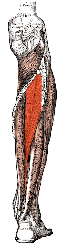
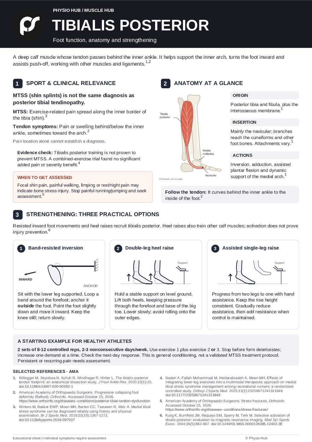
The tibialis posterior, also commonly called the posterior tibialis, is a deep calf muscle whose tendon passes behind the inner ankle and attaches to several bones of the foot. It helps support the medial longitudinal arch, turns the foot inward, and assists with plantar flexion, the movement used to point the foot downward.[1,2]
For athletes, understanding this muscle helps connect foot anatomy with practical training. The aim is to develop a foot that can accept load and contribute to push-off, while recognizing when shin or inner-ankle pain needs assessment.
Function and Sports Relevance
Supporting the foot during movement
Tibialis posterior contributes to arch support and helps the foot become a stable base for push-off. It works alongside other muscles, tendons, and ligaments; the arch does not depend on one muscle alone. These functions matter during repeated running steps and during jumping, landing, and changes of direction.[1,2]
A low arch does not automatically mean that tibialis posterior is weak. In a study of 60 adults without pain or disease, participants with flatter arches showed greater tibialis posterior activity during part of walking. This may reflect compensation, but the study did not show that the extra activity caused injury. Foot appearance, muscle activity, strength, and symptoms are different findings.[3]
The practical goal is comfortable, adaptable movement and sufficient strength for the athlete’s task. A useful assessment asks what the athlete can do, how symptoms respond, and what has changed in training, rather than judging the foot by arch height alone.
Tibialis posterior and shin splints
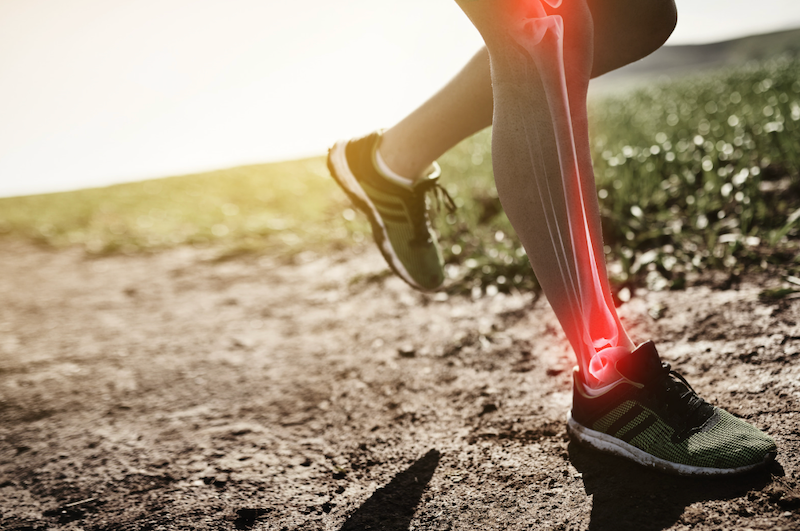
Medial tibial stress syndrome (MTSS), commonly called shin splints, is not the same diagnosis as posterior tibial tendinopathy. MTSS typically involves exercise-related pain and diffuse tenderness along the inner border of the tibia.[4]
Although tibialis posterior contributes to foot control, it should not be described as the sole cause of MTSS. Cadaver research challenges the simple explanation that traction from this muscle’s attachment accounts for the usual painful area. This does not exclude an indirect contribution to loading, but it makes “posterior tibialis shin splints” an imprecise label.[5]
| Condition | Typical presentation |
| MTSS | Exercise-related pain along the inner tibial border, with tenderness spread over a relatively broad area, commonly at least 5 cm.[4] |
| Posterior tibial tendon problem | Pain or swelling behind and below the inner ankle, sometimes extending toward the arch; push-off or heel raises may hurt.[2] |
| Tibial bone stress injury | More localized bony pain can occur; worsening symptoms may affect ordinary walking or produce pain at rest or at night.[6] |
These patterns help guide an examination. They cannot establish a diagnosis by themselves, and a broad area of tenderness does not rule out a bone stress injury.[4,6]
In a 2025 trial involving 40 recreational runners with MTSS, adding a lower-leg exercise program to other treatments improved some foot-posture measures and physical quality of life, but did not provide a statistically significant additional improvement in pain or MTSS severity. The program was not isolated tibialis posterior training, and the trial did not test prevention.[7]
Tibialis posterior strengthening can therefore be included in a broader foot and lower-leg program. It should not be advertised as a proven way to prevent or cure MTSS on its own.
When to seek assessment
Arrange an assessment if pain persists or repeatedly returns, if swelling develops along the inner ankle, or if the arch appears to be progressively changing. Difficulty performing a heel raise is useful information, but it does not identify the injured structure by itself.[2]
Stop painful impact exercise and seek prompt assessment for sharply localized shin pain, pain with normal walking, limping, or rest/night pain. These can be signs of a bone stress injury. Athletes and parents should not assume persistent shin pain is harmless “growing pain” or something to run through.[6]
Anatomy of the Tibialis Posterior
Origin
Posterior tibial and fibular surfaces and the interosseous membrane.[1]
Insertion
The navicular is a major attachment. Tendon slips also attach to the cuneiforms and other midfoot structures, commonly including the bases of the second to fourth metatarsals. The precise attachment pattern varies between individuals.[1,8]
Primary Actions
Inversion, turning the sole inward; adduction, moving the forefoot inward; assistance with plantar flexion; and dynamic support of the medial longitudinal arch.[1]
Its broad insertion links tibialis posterior to several parts of the foot, explaining why its role extends beyond lifting a single point of the arch.[1,8]
What the exercise evidence can tell us
A small MRI study found that resisted foot adduction recruited tibialis posterior relatively selectively, while single-leg heel raises recruited it together with other lower-leg muscles.[9] A 2026 study of 10 healthy adults also found high tibialis posterior activity during a resisted plantarflexion-inversion towel slide and unilateral heel raises.[10]
Muscle activation alone does not establish strength gains, symptom relief, or injury prevention. The instructions below are practical adaptations, not exact laboratory protocols. The 2026 paper was online as a journal pre-proof when this article was prepared.
These are general conditioning examples. If you currently have shin or inner-ankle pain, an assessment can help determine which exercises and loading levels are appropriate.
Effective Exercises for the Tibialis Posterior
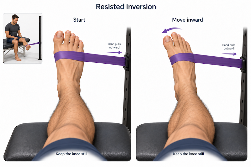
Exercise 1 | Resisted inversion with slight plantar flexion
Purpose: Apply resistance to inward foot movement, with the foot pointed slightly downward. This targets a major action of tibialis posterior, although other muscles also contribute.[1,9]
- Sit with your lower leg supported and your foot free to move.
- Loop a resistance band around the forefoot and anchor it toward the outside of the exercising foot so that it pulls the foot outward.
- Point the foot slightly downward, then move the forefoot inward against the band.
- Keep the knee and hip still. Return slowly, without letting the band pull the foot outward suddenly.
Technique cues: Keep the toes relaxed, use a comfortable range, and avoid turning the whole leg. Begin with resistance that allows smooth movement. Progress the band resistance when you can maintain the same control throughout the set.
Related option: For a seated inward foot slide, keep the same band pulling the forefoot outward. Place the forefoot on a small towel on a smooth floor, keep the heel planted and knee still, and slide the forefoot inward against the band. This is an alternative loading option, not a separately proven superior exercise.[9,10]
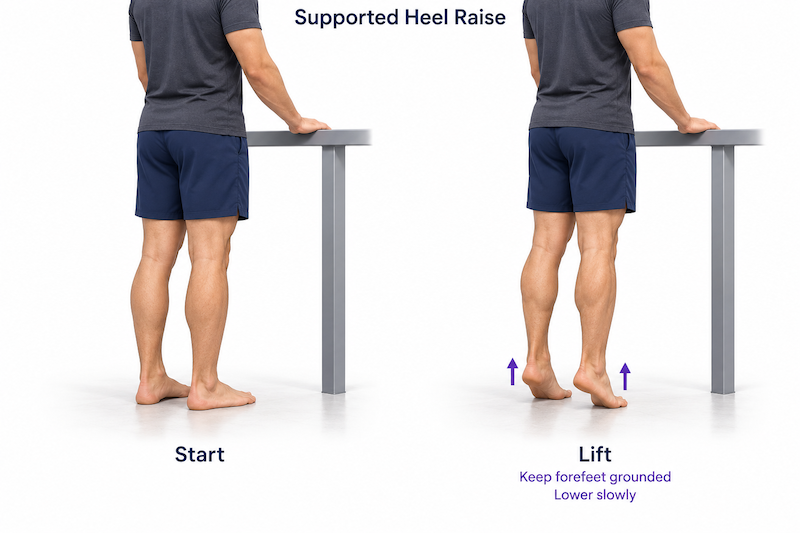
Exercise 2 | Supported double-leg heel raise
Purpose: Train the calf and foot-support muscles together. Heel raises load tibialis posterior, but they do not isolate it.[9]
- Stand on level ground with both feet in a comfortable position and hold a stable support lightly.
- Rise onto the balls of the feet, maintaining pressure through the base of the big toe and the rest of the forefoot.
- Lower smoothly and repeat without bouncing.
Technique cues: Avoid gripping the toes or shifting all your weight onto the outside edges of the feet. A small natural inward movement of the heel can occur; do not force the ankle into a rolled-out position. Start on the floor before considering the extra range available from a step.
Does toe position matter? In 14 healthy men, heel raises with the feet turned inward 30 degrees increased tibialis posterior activity compared with the other tested positions. This was an activation study, not proof of better treatment results. A comfortable neutral position remains a sensible starting choice; a forced toe-in stance is unnecessary.[11]
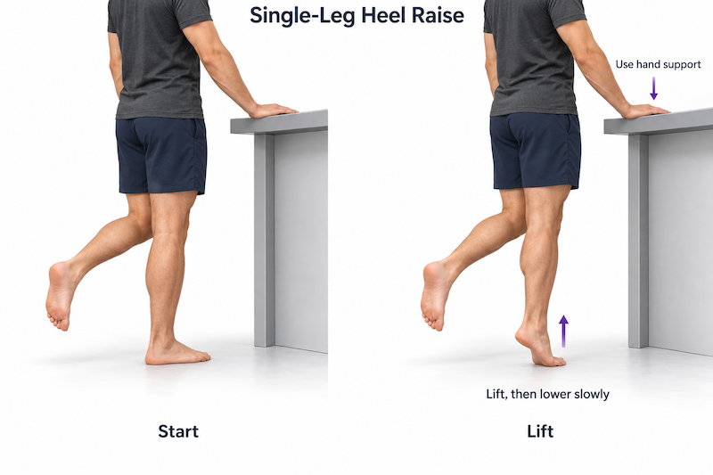
Exercise 3 | Assisted then loaded single-leg heel raise
Purpose: Increase the demand once double-leg heel raises are well controlled. Single-leg heel raises produced substantial tibialis posterior recruitment in laboratory studies.[9,10]
- Stand on one leg beside a stable support, using enough hand assistance to maintain balance and movement quality.
- Raise the heel through a comfortable range, then lower under control.
- Keep the forefoot supported and maintain a consistent heel-rise height from one repetition to the next.
Progression: Reduce hand assistance, then add external resistance, such as a dumbbell or a loaded backpack, as appropriate. If a full single-leg rise is too difficult, rise with both feet and shift somewhat more weight to one side for the lowering phase. This is a loading modification, not evidence that eccentric-only exercise is always better.
A heel raise challenges several muscles, balance, and tolerance to load. Repetition count alone should not be used to diagnose tibialis posterior weakness or decide whether an athlete is ready to return to sport.
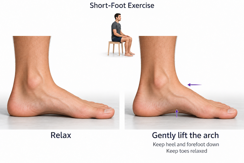
Exercise 4 | Short-foot control with standing integration
Purpose: Add whole-foot control to the strengthening program. This is a complementary exercise involving the intrinsic foot muscles; it is not selective tibialis posterior training.
- Sit with the heel, base of the big toe, and base of the little toe resting on the floor.
- Gently draw the ball of the foot toward the heel to create a small arch, without curling the toes or rolling onto the outer edge.
- Relax, repeat, and then practise the same gentle control in standing or during a slow supported single-leg task.
A 2024 trial in people with flexible flatfoot found benefits in selected foot-posture and balance measures from a program combining tibialis posterior strengthening and short-foot exercises. Because the exercises were combined, the results do not identify the effect of either one alone, and they do not establish injury prevention.[12]
How to choose the load and progress
The following is a practical training framework, not a validated tibialis-posterior-specific protocol. For a healthy athlete, two or three nonconsecutive strengthening sessions per week can be a reasonable starting schedule. Choose a resistance that feels challenging while allowing smooth movement, relaxed toes, and consistent heel-rise height. Stop a set before compensation becomes the main way to finish it.
Increase one demand at a time, such as resistance, repetitions, or single-leg loading. Monitor the response later that day and the following morning. Increasing pain, swelling, limping, or a lasting symptom increase is a reason to reduce the load and reassess. Do not use a tendinopathy “acceptable pain” rule to self-manage undiagnosed shin pain or suspected bone stress injury.
Once basic strength is established, progress toward the demands of the sport: repeated calf loading, balance during movement, landing, hopping, running, and changes of direction. An athlete recovering from injury should progress impact work according to the diagnosis and functional assessment, rather than simply completing a certain number of heel raises.
What if the posterior tibial tendon is already painful
Progressive resistance can be part of conservative management, but results vary. A 2009 trial of 36 adults with early tendon dysfunction found additional benefits from resistance exercise within an orthoses-and-stretching program.[13] A 2015 trial found that adding a moderate home strengthening program produced little extra benefit over orthoses and stretching.[14]
These studies involved people with tendon dysfunction, not healthy runners or athletes with MTSS. Exercise choice and loading should therefore be individualized. Footwear changes, orthoses, and temporary activity modification may also be appropriate when guided by assessment.[2,13,14]
< Take-Home Message >
Tibialis posterior supports the arch and foot control. Train it with resisted inward foot movement and progressive heel raises, supported by whole-foot exercises. Build strength across the lower leg, and have persistent shin or inner-ankle pain assessed.
< PDF file>
< Reference >
- Willegger M, Seyidova N, Schuh R, Windhager R, Hirtler L. The tibialis posterior tendon footprint: an anatomical dissection study. J Foot Ankle Res. 2020;13(1):25. doi:10.1186/s13047-020-00392-1
- American Academy of Orthopaedic Surgeons. Progressive collapsing foot deformity (flatfoot). OrthoInfo. Accessed October 25, 2026. https://www.orthoinfo.org/diseases–conditions/posterior-tibial-tendon-dysfunction
- Murley GS, Menz HB, Landorf KB. Foot posture influences the electromyographic activity of selected lower limb muscles during gait. J Foot Ankle Res. 2009;2:35. doi:10.1186/1757-1146-2-35
- Winters M, Bakker EWP, Moen MH, Barten CC, Teeuwen R, Weir A. Medial tibial stress syndrome can be diagnosed reliably using history and physical examination. Br J Sports Med. 2018;52(19):1267-1272. doi:10.1136/bjsports-2016-097037
- Stickley CD, Hetzler RK, Kimura IF, Lozanoff S. Crural fascia and muscle origins related to medial tibial stress syndrome symptom location. Med Sci Sports Exerc. 2009;41(11):1991-1996. doi:10.1249/MSS.0b013e3181a6519c
- American Academy of Orthopaedic Surgeons. Stress fractures. OrthoInfo. Accessed October 25, 2026. https://www.orthoinfo.org/diseases–conditions/stress-fractures/
- Naderi A, Fallah Mohammadi M, Heidaralizadeh A, Moen MH. Effects of integrating lower-leg exercises into a multimodal therapeutic approach on medial tibial stress syndrome management among recreational runners: a randomized controlled study. Orthop J Sports Med. 2025;13(2):23259671241311849. doi:10.1177/23259671241311849
- Uchiyama I, Edama M, Yokota H, et al. Anatomical study of sites and surface area of the attachment region of tibial posterior tendon attachment. Int J Environ Res Public Health. 2022;19(24):16510. doi:10.3390/ijerph192416510
- Kulig K, Burnfield JM, Requejo SM, Sperry M, Terk M. Selective activation of tibialis posterior: evaluation by magnetic resonance imaging. Med Sci Sports Exerc. 2004;36(5):862-867. doi:10.1249/01.MSS.0000126385.12402.2E
- Watson J, Eshenaur J, McClinton S. Electromyographic investigation of exercises that strengthen the foot’s arch. Journal of Biomechanics Open. Published online September 9, 2026. doi:10.1016/j.jbmo.2026.100026
- Akuzawa H, Imai A, Iizuka S, Matsunaga N, Kaneoka K. The influence of foot position on lower leg muscle activity during a heel raise exercise measured with fine-wire and surface EMG. Phys Ther Sport. 2017;28:23-28. doi:10.1016/j.ptsp.2017.08.077
- Kirmizi M, Sengul YS, Akcali O, Angin S. Effects of foot exercises and customized arch support insoles on foot posture, plantar force distribution, and balance in people with flexible flatfoot: a randomized controlled trial. Gait Posture. 2024;113:106-114. doi:10.1016/j.gaitpost.2024.05.030
- Kulig K, Reischl SF, Pomrantz AB, et al. Nonsurgical management of posterior tibial tendon dysfunction with orthoses and resistive exercise: a randomized controlled trial. Phys Ther. 2009;89(1):26-37. doi:10.2522/ptj.20070242
- Houck J, Neville C, Tome J, Flemister A. Randomized controlled trial comparing orthosis augmented by either stretching or stretching and strengthening for stage II tibialis posterior tendon dysfunction. Foot Ankle Int. 2015;36(9):1006-1016. doi:10.1177/1071100715579906