Physio Hub:
Evidence-Based Article
What Do X-Rays and MRIs Really Tell Us?
How to Use Medical Imaging Without Letting the Image Become the Entire Diagnosis
Key Points
- X-rays, CT scans, and MRIs are extremely valuable tools for identifying structural pathology.
- An abnormal imaging finding does not automatically identify the source of a person’s pain.
- Disc degeneration, cartilage defects, meniscal tears, and other structural findings can also occur in people who have no symptoms.
- Imaging can show what structures look like, but it usually cannot directly tell us why a particular activity hurts or how well someone can function.
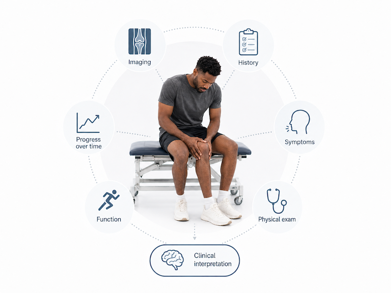
- The best interpretation combines imaging, history, symptoms, physical examination, function, and change over time.
Imaging Is Important—but It Is Not the Entire Answer
Imagine being told after an X-ray or MRI:
“Your joint space is almost gone.”
“You have degeneration.”
“There is a disc bulge.”
“You are bone on bone.”
It is understandable to immediately think:
“That must be why I hurt.”
Sometimes the structural finding is highly relevant.
Imaging is essential for identifying and characterizing many important conditions, including fractures, dislocations, tumors, infections, significant tissue injuries, and other structural pathology.
But CT and MRI have also been described as the “Donut of Truth,” reflecting the expectation that putting a patient through the scanner will reveal the answer to a difficult clinical problem.¹
The reality is more nuanced.
Imaging provides important information. It does not replace clinical reasoning.
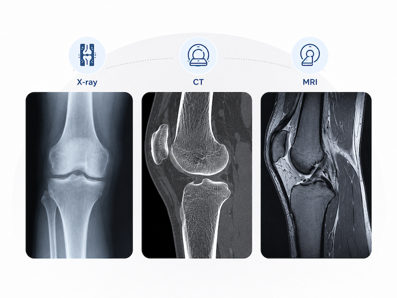
What Do X-Ray, CT, and MRI Show?
Different imaging methods answer different questions.
X-Ray
X-rays are particularly useful for assessing:
- Fractures
- Alignment
- Joint-space changes
- Osteophytes
- Deformity
- Many features of osteoarthritis
CT
CT provides more detailed visualization of bone and can be particularly useful for:
- Complex fractures
- Bone morphology
- Joint surfaces
- Bony alignment
MRI
MRI provides detailed information about many soft-tissue and intra-articular structures, including:
- Ligaments
- Tendons
- Muscles
- Menisci and labrum
- Cartilage
- Bone marrow
- Intervertebral discs
- Neural and surrounding soft tissues
These are extremely useful capabilities.
What routine static imaging does not directly measure, however, is:
- Strength
- Power
- Balance
- Load tolerance
- Running ability
- Sport performance
- Confidence
- Whether a particular movement is tolerable
Those questions usually require functional assessment.
An Imaging “Abnormality” Does Not Automatically Equal Pain
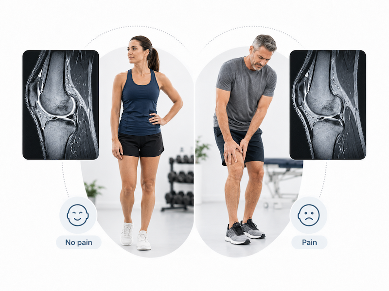
One of the most important lessons from imaging research is that structural abnormalities are also found in people without symptoms.
A systematic review involving 3,110 asymptomatic individuals found lumbar disc degeneration in approximately 37% of people at age 20, increasing to 96% by age 80.²
Disc bulges were found in approximately 30% of asymptomatic 20-year-olds and 84% of asymptomatic 80-year-olds.²
The knee provides another example.
A systematic review of 4,751 asymptomatic adults with no previous knee injury found MRI evidence of:
- Cartilage defects in approximately 24%
- Meniscal tears in approximately 10%
- Bone marrow lesions in approximately 18%
- Osteophytes in approximately 25%
with several findings becoming considerably more common with age.³
Therefore:
Seeing a structural finding on a scan does not prove that the finding is responsible for the patient’s symptoms.
Does That Mean Imaging Findings Do Not Matter?
No.
That would be an equally problematic interpretation.
Fractures, severe joint disease, major structural injuries, tumors, infection, and many other findings can clearly be clinically important.
Even in osteoarthritis, greater structural severity can be associated with greater symptoms.
The important point is that the relationship is imperfect.
A systematic review examining knee osteoarthritis found substantial variability between radiographic OA and knee pain: some people had radiographic disease without pain, while others had knee pain without corresponding radiographic OA.⁴
The appropriate conclusion is therefore not:
“Ignore the scan.”
It is:
“Interpret the scan in the context of the person.”
More Imaging Does Not Automatically Mean Better Decisions
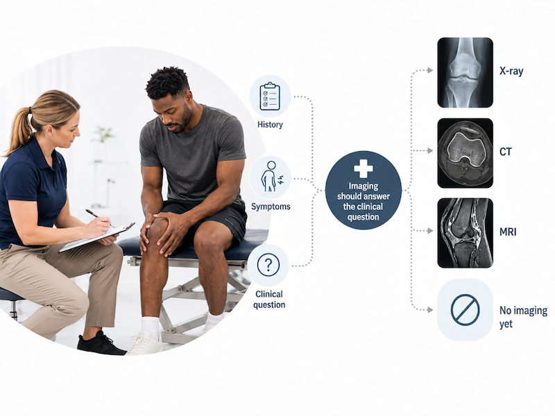
A more advanced scan is not always a better scan for the question being asked.
In one study, 41 spine surgeons evaluated 30 thoracolumbar fractures.
Using radiographs alone, 43.4% of fractures were classified correctly. Adding CT substantially improved classification accuracy. Adding MRI produced only a small additional improvement, and the decision regarding surgical fixation did not significantly change after MRI was added.⁵
This does not mean MRI has no role in spinal trauma.
It illustrates a broader principle:
The appropriate imaging study depends on the clinical question you are trying to answer.
“Just Get an MRI” Is Not Always Better Care
Imaging can be invaluable when there is a clear indication.
But routine imaging in situations where serious pathology is not suspected does not necessarily improve outcomes.
A systematic review and meta-analysis of randomized trials found that immediate routine lumbar imaging for patients with low back pain without indicators of serious underlying disease did not improve pain or function compared with usual care without immediate imaging.⁶
The question should therefore not simply be:
“Can we get an MRI?”
A better question is:
“What clinical question will this imaging help us answer, and will the result change management?”
The Language Used to Explain Imaging Matters
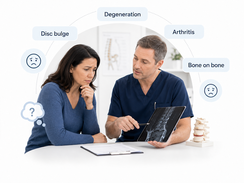
Terms such as:
- “Degeneration”
- “Arthritis”
- “Disc bulge”
- “Bone on bone”
can sound frightening.
A randomized scenario-based study involving people with and without low back pain found that labels such as “disc bulge,” “degeneration,” and “arthritis” increased perceived need for imaging, surgery, and second opinions and were associated with lower recovery expectations compared with more neutral labels.⁷
This does not mean clinicians should hide imaging findings or minimize important pathology.
It means imaging should be communicated accurately and in context, without unnecessarily convincing patients that their bodies are fragile or damaged beyond repair.
What Should Rehabilitation Assess Alongside Imaging?
Rehabilitation is concerned not only with structure, but also with function.
Relevant questions may include:
- What activities reproduce the symptoms?
- What is the available range of motion?
- What are the person’s strength and power capacities?
- How well can they balance and control movement?
- What loads can they currently tolerate?
- What sport, work, or daily activities are limited?
- How are symptoms changing over time?
- How does the person respond to progressive exercise or treatment?
A qualitative study of expert musculoskeletal physical therapists found that imaging was interpreted as part of a broader clinical reasoning process that also considered functional consequences, precautions, and the need for referral rather than being treated as a stand-alone answer.⁸
A useful distinction is:
Imaging can help tell us what is structurally present.
Functional assessment helps tell us what that finding means for this person.
What We Know
Current evidence supports several important conclusions:
- Imaging is highly valuable when used to answer an appropriate diagnostic or management question.
- Degenerative and structural imaging findings are common in asymptomatic people.²˒³
- Structural severity and symptoms may be related, but the relationship is not one-to-one.⁴
- More or earlier imaging does not automatically produce better patient outcomes.⁶
- Imaging findings should be interpreted alongside clinical and functional information.
What We Still Don’t Know
There are still questions that imaging alone cannot answer reliably:
- Which of several imaging abnormalities is contributing most to one individual’s pain?
- Whether an asymptomatic structural finding will eventually become symptomatic
- Exactly how much pain or disability an individual will experience based on a scan
- How much structural change represents harmless adaptation or aging versus a clinically meaningful problem in every individual
This uncertainty is one reason clinical reasoning remains necessary.
What Athletes, Parents, Coaches, and Trainers Should Know
If an imaging report identifies an abnormality, do not automatically conclude:
“My body is damaged.”
“I can’t play anymore.”
“This finding explains everything.”
But do not dismiss the imaging either.
Ask instead:
Does this finding fit the symptoms?
Does it change treatment or precautions?
What does the person’s functional assessment show?
How is the condition changing over time?
The value of the scan comes from understanding what it means for the individual, not simply from the words written in the report.
< Take-Home Message >
X-rays, CT scans, and MRIs are powerful and essential tools in modern healthcare.
But:
An image is a piece of clinical information—not the entire clinical answer.
An abnormal scan does not automatically mean someone should be in pain, and a relatively normal scan does not mean that meaningful symptoms or functional limitations cannot exist.
Good decision making combines:
Imaging + History + Symptoms + Physical Examination + Function + Change Over Time
The goal is not to believe or disbelieve imaging.
The goal is to understand what the imaging means and use it appropriately.
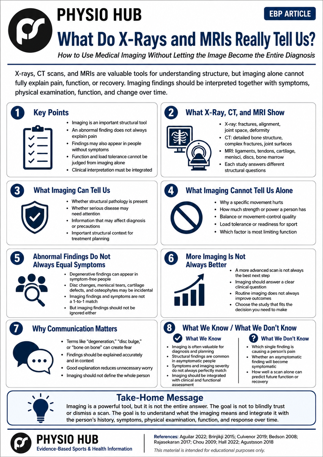
<PDF file>
Read More



< Reference >
- Aguilar C, Gunderman R. The donut of truth. J Am Coll Radiol. 2022;19(8):992-993. doi:10.1016/j.jacr.2022.04.004.
- Brinjikji W, Luetmer PH, Comstock B, et al. Systematic literature review of imaging features of spinal degeneration in asymptomatic populations. AJNR Am J Neuroradiol. 2015;36(4):811-816. doi:10.3174/ajnr.A4173.
- Culvenor AG, Øiestad BE, Hart HF, Stefanik JJ, Guermazi A, Crossley KM. Prevalence of knee osteoarthritis features on magnetic resonance imaging in asymptomatic uninjured adults: a systematic review and meta-analysis. Br J Sports Med. 2019;53(20):1268-1278. doi:10.1136/bjsports-2018-099257.
- Bedson J, Croft PR. The discordance between clinical and radiographic knee osteoarthritis: a systematic search and summary of the literature. BMC Musculoskelet Disord. 2008;9:116. doi:10.1186/1471-2474-9-116.
- Rajasekaran S, Vaccaro AR, Kanna RM, et al. The value of CT and MRI in the classification and surgical decision-making among spine surgeons in thoracolumbar spinal injuries. Eur Spine J. 2017;26(5):1463-1469. doi:10.1007/s00586-016-4623-0.
- Chou R, Fu R, Carrino JA, Deyo RA. Imaging strategies for low-back pain: systematic review and meta-analysis. Lancet. 2009;373(9662):463-472. doi:10.1016/S0140-6736(09)60172-0.
- Hall AM, Ferreira PH, Maher CG, et al. Effect of diagnostic labelling on management intentions for non-specific low back pain: a randomized scenario-based experiment. Eur J Pain. 2022;26(9):1913-1924.
- Agustsson H. Diagnostic Musculoskeletal Imaging: How Physical Therapists Utilize Imaging in Clinical Decision-Making. Doctoral dissertation. Nova Southeastern University; 2018.
Stay Informed with Physio Hub
Unlock a world of knowledge with our expertly curated articles on fitness, rehabilitation, and health & diet. Stay ahead with the latest evidence-based insights and practical advice. Subscribe now to receive updates and never miss out on new content!