Physio Hub:
Evidence-Based Article
Patellofemoral Pain Syndrome in Athletes
Patellofemoral pain syndrome (PFPS), often referred to simply as patellofemoral pain (PFP) in contemporary literature, is a clinical anterior-knee-pain condition characterized by peri- or retropatellar pain during activities that load the patellofemoral joint in knee flexion.¹⁻³
Typical provoking activities include:
- Squatting
- Stair ascent or descent
- Running
- Jumping and landing
- Prolonged sitting¹⁻³
PFPS is best understood as a multifactorial, load-related knee pain condition, rather than a disorder produced by one abnormal structure or movement pattern.¹⁻⁴
What Is Patellofemoral Pain Syndrome?
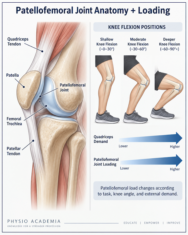
The patellofemoral joint is formed by the patella and femoral trochlea.
During knee flexion and extension, the patella moves within the trochlea while transmitting forces generated by the quadriceps mechanism.
Activities requiring greater knee flexion and quadriceps demand can substantially increase patellofemoral loading.
In an athlete with PFPS, symptoms may develop when the demands being placed on the patellofemoral system exceed the athlete’s current ability to tolerate those demands.
Potential contributors include:
- Excessive or rapidly increased loading
- Dynamic knee valgus
- Hip or quadriceps dysfunction
- Trunk-control deficits
- Altered patellar mechanics
- Foot mechanics
- Mobility restrictions¹⁻⁴
However, no single mechanism explains every case.
PFPS should therefore not automatically be reduced to “patellar maltracking,” VMO weakness, or one biomechanical abnormality.
Typical Symptoms
Athletes commonly report:
- Peripatellar or retropatellar pain
- Pain with squatting
- Pain with stairs
- Running-related pain
- Pain with jumping or landing
- Pain after prolonged sitting
- Symptoms that increase with training volume¹⁻³
The clinical pattern is primarily one of pain during loaded knee flexion.
Why Is It Important in Athletes?
PFP is common in runners and athletes participating in jumping, cutting, and pivoting sports.²,³
Female athletes are affected more frequently in many populations, although the difference cannot be explained by one isolated anatomical or biomechanical factor.
PFPS can affect:
- Running volume
- Jump tolerance
- Squatting and lunging
- Training participation
- Competition
- Long-term sport participation³,⁴
The condition should not always be considered a minor, rapidly resolving knee complaint.
Persistent symptoms occur in a meaningful proportion of patients.
For athletes, the rehabilitation objective is therefore not simply to reduce anterior knee pain, but to restore repeated patellofemoral loading capacity for sport.
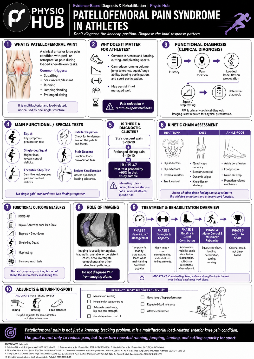
Functional Diagnosis
PFPS is primarily a clinical diagnosis.¹,⁴
Imaging is not required to confirm a typical presentation.
Functional diagnosis should integrate:
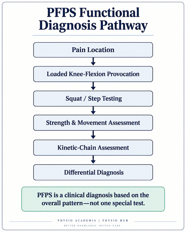
history + pain location + load provocation + functional testing + differential diagnosis.
One of the most important findings is reproduction of familiar peri- or retropatellar pain during a task that loads the knee in flexion.
Step 1: Identify the Symptom Pattern
The history should determine:
- Location of pain
- Duration and onset
- Recent changes in training volume
- Changes in running or jumping exposure
- Which movements provoke symptoms
- Pain during stair ascent or descent
- Pain with prolonged sitting
- Response during and after training
The clinician should look for a coherent load-related anterior knee pain pattern rather than relying on one named special test.
Step 2: Reproduce Symptoms With Loaded Knee Flexion
Next, the clinician should determine whether the athlete’s familiar anterior knee pain is reproduced during tasks that load the patellofemoral joint in knee flexion.
Common examples include:
- Squatting
- Single-leg squatting
- Step-down tasks
- Stair descent
- Running
- Jumping
- Landing
- Cutting
The key finding is not simply that the knee hurts.
The important question is whether familiar peri- or retropatellar pain is reproduced during loaded knee-flexion tasks.
In athletes, lower-demand tasks may not reproduce symptoms even when higher-load or higher-speed activities do. Therefore, testing should eventually reflect the athlete’s sport demands.
Step 3: Consider Differential Diagnosis
PFPS is a clinical diagnosis, but not all anterior knee pain is PFPS.
Depending on the athlete’s age, symptom location, trauma history, swelling, instability, locking, and irritability, clinicians should consider other possible conditions such as:
- Patellar tendinopathy
- Osgood-Schlatter disease
- Sinding-Larsen-Johansson syndrome
- Meniscal pathology
- Patellar instability
- Fat pad irritation
- Plica syndrome
- Osteochondral lesion
- Ligament injury
- Referred pain from the hip or lumbar spine
Clinicians should be especially cautious when symptoms include:
- Clear traumatic onset
- Significant swelling
- Locking
- Giving way
- Night pain
- Progressive symptoms
- Symptoms that do not match a typical PFPS pattern
Functional diagnosis should confirm that the clinical pattern is consistent with PFPS while also identifying findings that may suggest another condition.
Squatting as a Key Functional Test
Squatting is one of the most clinically useful symptom-provocation tasks for PFP.¹,⁶
Evaluation may include:
- Reproduction of familiar anterior knee pain
- Depth at which pain develops
- Response to increasing load
- Trunk strategy
- Hip control
- Knee control
- Side-to-side differences
A single-leg squat can increase the strength and neuromuscular demands and may reveal findings that are not apparent during bilateral squatting.
Main Functional and Special Tests
There is no single gold-standard physical test for PFPS.¹,⁵
Tests should therefore be interpreted together.
Eccentric Step Test
The eccentric step test challenges the knee during loaded flexion and eccentric control.
In one diagnostic-accuracy study, it demonstrated a sensitivity of 0.82, the highest among the individual tests evaluated.⁵
It also allows observation of:
- Hip control
- Dynamic valgus
- Trunk mechanics
- Eccentric knee control
- Symptom response
Patellar Palpation
Palpation around the patella can determine whether familiar symptoms are reproduced.
The same diagnostic study reported a sensitivity of 0.81 for palpation.⁵
Tenderness alone, however, does not establish the diagnosis.
Stair Descent
Stair descent places substantial eccentric demand on the quadriceps and increases patellofemoral loading.
It can therefore be a particularly useful real-world provocation test.
Patellar Tilt and Mobility
Patellar tilt and mobility may provide additional information regarding local mechanics.
These tests are best used as adjunct findings, not as proof that patellar alignment is the cause of the athlete’s pain.
Patellar Compression / Grind Testing
Compression or grind tests have traditionally been used during the patellofemoral examination.
A positive result should not be interpreted as a definitive diagnosis of PFPS.
History and functional pain provocation are more important components of the overall clinical pattern.
Resisted Knee Extension
Resisted knee extension can assess:
- Symptom reproduction
- Quadriceps loading tolerance
- Strength deficits
- Side-to-side differences
It is especially useful when progressing from diagnosis toward functional rehabilitation planning.
Is There a Diagnostic Cluster?
There is no universally validated diagnostic cluster for athletes with PFPS.
One diagnostic-accuracy study found that the combination of:
pain rated 3-10/10 during stair descent
plus
pain rated 6-10/10 during prolonged sitting
produced a positive likelihood ratio of 19.47 and approximately 95% post-test probability in that study population.⁵
This is an interesting rule-in finding, but it should be interpreted carefully.
It came from a single diagnostic study and should not be treated as a universally validated athlete-specific clinical prediction rule.
A more robust clinical approach remains:
typical pain location + pain during loaded knee flexion + reproducible symptoms during squat/step tasks + functional examination + exclusion of competing diagnoses
Evaluate the Kinetic Chain
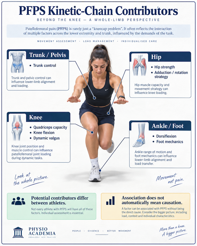
For an athlete, functional diagnosis should also investigate why sport demands may be exceeding the current capacity of the patellofemoral system.
Potential areas to assess include:
Hip and Trunk
- Hip abductor strength
- Hip extensor strength
- Hip external-rotator strength
- Trunk control
Knee
- Quadriceps capacity
- Eccentric control
- Knee-flexion strategy
- Dynamic valgus
Ankle and Foot
- Ankle dorsiflexion
- Foot posture
- Navicular drop
- Relevant pronation mechanics¹⁻⁴
Importantly, finding dynamic valgus does not prove that valgus is causing the pain.
The impairment should be linked to the athlete’s symptoms and meaningful sport function before it becomes a rehabilitation target.
Functional Outcome Measures
Useful patient-reported measures include:
KOOS-PF
A patellofemoral-specific patient-reported outcome measure.⁶
Kujala / Anterior Knee Pain Scale
A widely used measure of pain and function in patellofemoral disorders.⁶
Functional monitoring can also include:
- Squat
- Step-up
- Step-down
- Single-leg squat
- Hop testing
- Balance/reach testing
A 2026 secondary analysis found that changes in step-up and step-down performance were more closely related to patient-reported improvement than changes in the squat test alone.⁷
This highlights an important point:
the best test for provoking symptoms at diagnosis may not necessarily be the best test for monitoring recovery.
Role of Imaging
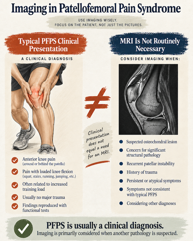
Typical PFPS is primarily a clinical diagnosis, and imaging is not routinely required.¹,³,⁴
Imaging becomes more relevant when there is concern for:
- Osteochondral pathology
- Significant structural injury
- Patellar instability
- Trauma
- Atypical or persistent symptoms
Imaging should generally be used to investigate alternative or concomitant pathology rather than to “confirm” uncomplicated PFPS. Expert clinicians have similarly emphasized that diagnosis is clinical and imaging should mainly screen for other pathology.⁴
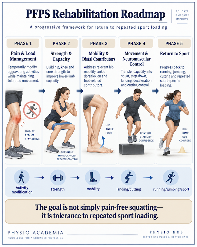
Treatment and Rehabilitation Overview
**Conservative rehabilitation is first-line management.**¹,³,⁴
The central framework is:
education + activity/load modification + progressive exercise therapy.
Phase 1: Pain and Load Management
Identify the activities currently exceeding the athlete’s patellofemoral capacity.
Examples may include:
- Increased running mileage
- Hills
- High jump volume
- Deep squatting
- Repetitive stairs
- Cutting volume
The goal is usually not complete rest.
Training should be temporarily modified to reduce excessive symptom provocation while maintaining tolerable activity.
Phase 2: Strength and Capacity
Current evidence generally supports rehabilitation addressing the hip and knee, often with trunk/core considerations, rather than relying on isolated quadriceps strengthening alone.¹,³,⁴
Treatment should still be individualized according to examination findings.
A 2025 randomized clinical trial found that functional strength training produced greater improvements in pain and knee function than standard strength training and was accompanied by increased gluteus maximus activation and reduced knee valgus.⁸
Phase 3: Mobility and Distal Contributors
When clinically relevant, rehabilitation may also address:
- Hip mobility
- Ankle dorsiflexion
- Foot function
- Relevant soft-tissue restrictions
A 2026 randomized trial in male collegiate athletes found that adding hip- and ankle-mobility-based rehabilitation improved pain, Kujala scores, hip rotation, ankle dorsiflexion, and dynamic balance.⁹
For selected patients with relevant foot findings, adding short-foot exercise to hip- and knee-focused rehabilitation has also improved pain, function, and balance compared with hip and knee exercise alone.¹²
Phase 4: Motor Control and Functional Retraining
Strength alone may not automatically change sport movement.
The athlete must learn to express newly developed capacity during:
- Squatting
- Step-down
- Landing
- Deceleration
- Cutting
- Running
In active females with PFPS, valgus-control instruction improved pain, dynamic valgus, hip torque, and functional performance.¹⁰
A 2026 randomized trial in recreational sprinters also found greater improvements in pain, dynamic balance, and hop performance with dynamic proprioceptive training compared with conventional strengthening.¹¹
Phase 5: Return to Sport
Return to sport should be criteria based rather than time based.
Relevant considerations may include:
- Pain-free or acceptable squatting
- Stair tolerance
- Running tolerance
- Quadriceps capacity
- Hip and trunk capacity
- Step-down control
- Jump/hop performance
- Balance
- Repeated-load tolerance
- Athlete confidence³,¹³
The final question is not simply whether everyday pain has disappeared.
It is whether the athlete can repeatedly tolerate the specific intensity, volume, speed, and movement demands of sport.
Adjunctive Treatment
Patellar taping, bracing, and foot orthoses may be useful selectively to modify symptoms or make rehabilitation easier.¹,³,⁴
They are best considered adjuncts, rather than stand-alone treatments.
The primary long-term intervention remains progressive rehabilitation that restores the athlete’s physical and functional capacity.
< Summary >
Patellofemoral pain syndrome should not simply be viewed as a kneecap “tracking problem.”
It is better understood as a multifactorial, load-related anterior knee pain condition in which sport demands exceed the current capacity of the patellofemoral system and surrounding kinetic chain.
Functional diagnosis should integrate:
pain location → loaded knee-flexion provocation → squat/step testing → strength and movement assessment → kinetic-chain contributors → differential diagnosis.
There is no single definitive special test or universally validated athlete-specific diagnostic cluster.
Rehabilitation generally progresses through:
load management → hip/knee/trunk capacity → mobility when relevant → neuromuscular retraining → sport-specific loading.
For athletes, the ultimate goal is not merely a knee that is pain-free during one squat.
It is a knee and kinetic chain capable of repeatedly running, jumping, landing, cutting, and competing at the required sport load.
<PDF file>
Read More
< Reference >
- Callahan EA, Chin KE, Chu SK. Current evidence of evaluation and management of the athlete with patellofemoral pain syndrome. Curr Phys Med Rehabil Rep. 2025;13:30. doi:10.1007/s40141-025-00502-9.
- Petersen W, Rembitzki I, Liebau C. Patellofemoral pain in athletes. Open Access J Sports Med. 2017;8:143-154. doi:10.2147/OAJSM.S133406.
- Halabchi F, Abolhasani M, Mirshahi M, Alizadeh Z. Patellofemoral pain in athletes: clinical perspectives. Open Access J Sports Med. 2017;8:189-203. doi:10.2147/OAJSM.S127359.
- Curran AJ, Neal BS, Barber P, Bartholomew C, Morrissey D, Lack SD. Clinicians’ experience of the diagnosis and management of patellofemoral pain: a qualitative exploration. Musculoskelet Sci Pract. 2022;58:102530. doi:10.1016/j.msksp.2022.102530.
- Mostafaee N, Pashaei-Marandi M, Negahban H, et al. Examining the diagnostic accuracy of common physical examination and functional tests in the diagnosis of patellofemoral pain syndrome among patients with anterior knee pain. Physiother Theory Pract. 2024;40(4):843-855. doi:10.1080/09593985.2022.2158053.
- Chamorro-Moriana G, Espuny-Ruiz F, Ridao-Fernández C, Magni E. Clinical value of questionnaires & physical tests for patellofemoral pain: validity, reliability and predictive capacity. PLoS One. 2024;19(4):e0302215. doi:10.1371/journal.pone.0302215.
- de Morais Machado E, Matthews M, Rathleff MS, Serrão FV, Vicenzino B. Is outcome observed in performance of clinical tests associated with patient-reported outcome measures after patellofemoral pain treatment? Secondary analysis of data from a randomized clinical trial. Braz J Phys Ther. 2026;30(4):101593. doi:10.1016/j.bjpt.2026.101593.
- Xiong Z, Zheng W, Wang H, et al. Effects of functional strength training on pain, function, and lower extremity biomechanics in patients with patellofemoral pain syndrome: a randomized clinical trial. J Orthop Surg Res. 2025;20:50. doi:10.1186/s13018-025-05482-z.
- Xu H, Feng Z, et al. Effects of a 6-week hip and ankle mobility-based rehabilitation program on clinical, neuromuscular, and functional outcomes in male collegiate athletes with patellofemoral pain: a randomized controlled trial. Life (Basel). 2026;16(6):1013. doi:10.3390/life16061013.
- Emamvirdi M, Letafatkar A, Khaleghi Tazji M. The effect of valgus control instruction exercises on pain, strength, and functionality in active females with patellofemoral pain syndrome. Sports Health. 2019;11(3):223-237. doi:10.1177/1941738119837622.
- Devadharshini B, Subramanian SS, Thirumalai J, et al. Dynamic proprioceptive training improves functional recovery in sprinters with patellofemoral pain: a randomized trial. PLoS One. 2026;21(7):e0353107. doi:10.1371/journal.pone.0353107.
- Kamel AM, Ghuiba K, Abd Allah DS, Fayaz NA, Abdelkader NA. Effect of adding short foot exercise to hip and knee focused exercises in treatment of patients with patellofemoral pain syndrome: a randomized controlled trial. J Orthop Surg Res. 2024;19:207. doi:10.1186/s13018-024-04688-x.
- Vora M, Curry E, Chipman A, Matzkin E, Li X. Patellofemoral pain syndrome in female athletes: a review of diagnoses, etiology and treatment options. Orthop Rev (Pavia). 2018;9(4):7281. doi:10.4081/or.2017.7281.
- Hasan S, Alonazi A, Anwer S, et al. Efficacy of patellar taping and electromyographic biofeedback training at various knee angles on quadriceps strength and functional performance in young adult male athletes with patellofemoral pain syndrome: a randomized controlled trial. Pain Res Manag. 2022;2022:8717932. doi:10.1155/2022/8717932.
Stay Informed with Physio Hub
Unlock a world of knowledge with our expertly curated articles on fitness, rehabilitation, and health & diet. Stay ahead with the latest evidence-based insights and practical advice. Subscribe now to receive updates and never miss out on new content!