Physio Hub:
Evidence-Based Article
Lumbar Disc Herniation
Lumbar disc herniation (LDH) is a spinal disorder in which disc material protrudes or extrudes beyond its normal boundary and may irritate or compress nearby neural structures.¹,²
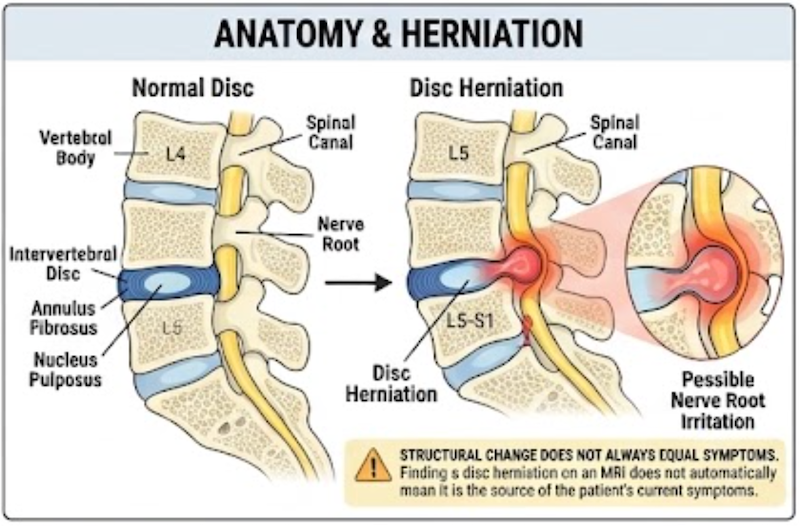
When neural structures are affected, athletes may experience low back pain, radiating leg pain, paresthesia, numbness, and sometimes motor deficits. L4-L5 and L5-S1 account for many symptomatic cases.
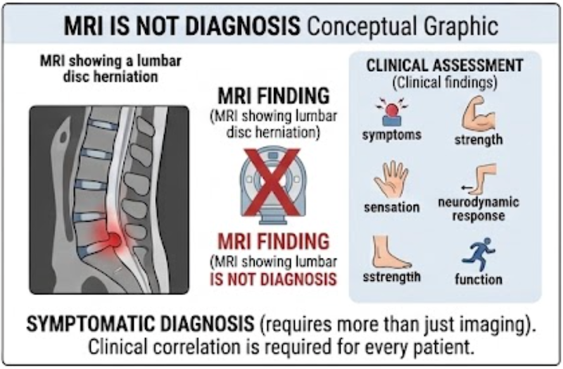
A critical clinical principle is that an MRI-confirmed disc herniation does not automatically prove that the disc finding is responsible for the athlete’s symptoms.
Diagnosis should integrate:
**symptoms + neurological findings + neurodynamic testing + imaging + function.**³⁻⁶
What Is Lumbar Disc Herniation?
The intervertebral disc sits between adjacent vertebral bodies and contributes to:
- Load distribution
- Shock absorption
- Spinal mobility
The inner nucleus pulposus is surrounded by the annulus fibrosus.
With cumulative mechanical loading, tissue changes, or trauma, disc material may protrude through or beyond the annular region and enter the spinal canal or neural foramen.¹,²
When the herniated material irritates or compresses a nerve root, radicular symptoms may develop.
Typical Symptoms
Clinical presentation may include:
- Low back pain
- Buttock pain
- Radiating leg pain
- Paresthesia
- Numbness
- Sensory loss
- Muscle weakness
- Reduced tolerance to athletic activity¹⁻³
However, symptoms and MRI findings do not always correlate perfectly.³,⁴
Therefore:
disc abnormality on MRI ≠ automatically the source of pain.
The imaging must make sense in the context of the athlete’s symptoms and examination.
Why Is It Important in Athletes?
Athletes may expose the lumbar spine to repeated:
- Flexion
- Compression
- Rotation
- High-load training
- Cumulative microtrauma⁷
Some younger athletes may also develop symptomatic disc herniation following a more distinct traumatic event.⁷
For athletes, the clinical challenge goes beyond reducing pain.
Sport may require repeated:
- Sprinting
- Jumping
- Rotation
- Throwing
- Heavy lifting
- Contact
- Repeated spinal loading
Therefore, the key question is not simply:
“Does the athlete feel better?”
It is:
“Can the athlete again tolerate the spinal and whole-body demands required for sport?”
Athletes with LDH and chronic low back pain have demonstrated impairments in measures including hip internal rotation, trunk flexibility, balance, and trunk endurance compared with pain-free athletes.⁸
Functional Diagnosis
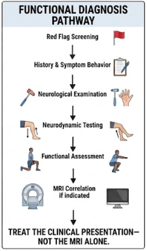
Functional diagnosis of lumbar disc herniation should determine whether the athlete’s clinical and neurological presentation corresponds to the suspected disc lesion.
There is currently no well-established athletic LDH diagnostic cluster in which a fixed number of positive special tests confirms the diagnosis.
Instead, assessment should integrate:
history → neurological examination → neurodynamic testing → functional assessment → MRI correlation.
The purpose is not only to determine whether a disc herniation is present.
It is also to determine whether the disc finding meaningfully relates to the athlete’s symptoms, neurological presentation, and functional limitations.
Screen for Red Flags First
Before proceeding with routine sports rehabilitation, clinicians must identify presentations requiring urgent medical evaluation.
Particular concern exists for possible cauda equina syndrome.
Potential warning findings include:
- Bowel dysfunction
- Bladder dysfunction
- Major sensory loss
- Major motor loss
- Rapidly progressive weakness
- Progressive loss of walking ability³
These presentations should not simply be managed as routine mechanical low back pain.
History and Symptom Behavior
The history should determine:
- Whether symptoms are confined to the back or radiate into the leg
- Presence of numbness or paresthesia
- Perceived weakness
- Positions or movements that aggravate symptoms
- Sport-specific symptom reproduction
- Acute versus gradual onset
- Previous trauma
- Whether neurological symptoms are stable, improving, or progressing
The combination of low back pain plus radiating neurological symptoms should increase suspicion of lumbar nerve-root involvement.
Neurological Examination
The neurological examination should include assessment of:
Motor Function
Manual muscle testing or other appropriate strength testing should identify meaningful motor deficits.
Sensory Function
Sensory changes and side-to-side differences should be assessed.
Gait and Functional Neurological Assessment
The clinician should determine whether neurological symptoms are affecting gait or basic functional ability.³
The purpose is not simply to find one weak muscle.
The examination should determine whether the athlete’s symptoms and neurological findings form a coherent pattern of nerve-root involvement.
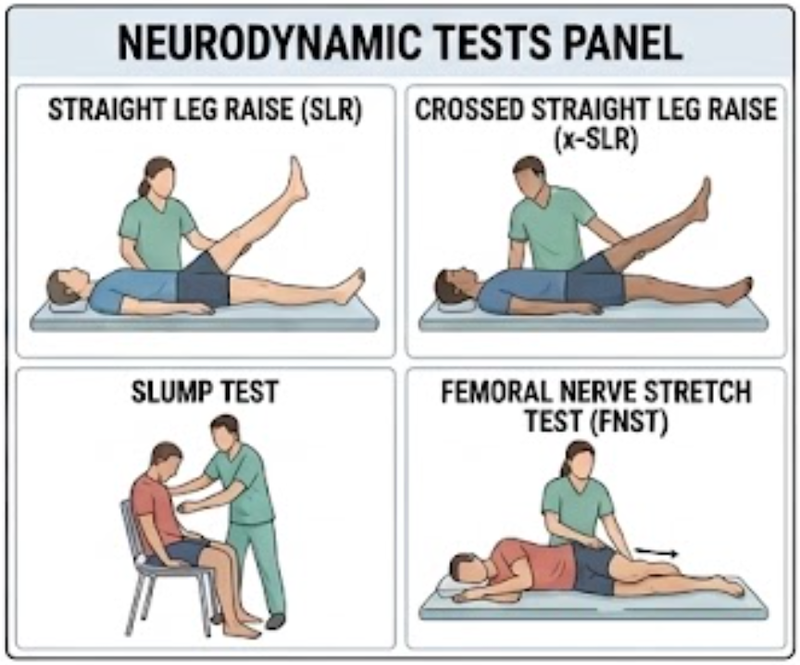
Main Special Tests
Straight Leg Raise
The straight leg raise (SLR) remains one of the most commonly used neurodynamic screens for lumbar radicular involvement.
With the athlete supine, the leg is progressively elevated and the clinician determines whether the patient’s familiar neurological symptoms are reproduced.
SLR is generally considered relatively sensitive, but its specificity is limited.⁵,⁹
In adolescent lumbar disc herniation, a positive SLR has been reported in approximately 90% of patients.⁷
Importantly:
limited hamstring flexibility alone does not make the SLR positive.
Reproduction of the patient’s relevant radicular symptoms is more meaningful.
Crossed Straight Leg Raise
During the crossed SLR, elevation of the unaffected leg reproduces symptoms in the symptomatic leg.
It is commonly used as an adjunct to SLR, although the evidence regarding reliability remains inconclusive.⁵
Slump Test
The slump test alters neural loading through combined spinal, hip, knee, and ankle positioning.
In one prospective comparison, the slump test demonstrated greater sensitivity than SLR for L4-L5 and combined L4-L5/L5-S1 disc herniation.¹⁰
It should still be interpreted as part of the full clinical examination rather than as proof of disc herniation by itself.
Femoral Nerve Stretch Test
Femoral nerve tension testing may be useful when higher lumbar nerve-root involvement is suspected.
Because lower lumbar disc levels are more common, it is generally less central than SLR but can be useful when the symptom distribution suggests more proximal involvement.
Extended Straight Leg Raise
Structural differentiation can be added to SLR using maneuvers such as:
- Ankle dorsiflexion
- Hip internal rotation
An MRI-referenced study reported a sensitivity of 0.85 for an extended SLR approach and a meaningful association with MRI-confirmed disc herniation.⁹
These differentiation maneuvers may help determine whether symptoms behave more like a neural response than simple posterior-chain muscle restriction.
Compression Overload Test
The Compression Overload Test is a newer proposed diagnostic test.
A 2025 MRI-validated cross-sectional study reported 92% sensitivity and 90.57% diagnostic accuracy, outperforming SLR in that particular sample.¹¹
However, this finding is still early.
The Compression Overload Test should not yet be considered a replacement for established neurological and neurodynamic examination, and further validation is needed.
Is There a Diagnostic Cluster?
At present, there is no sufficiently validated special-test cluster specific to lumbar disc herniation in athletes.
A clinically meaningful pattern instead combines:
- Radicular symptoms
- Neurological deficits
- Positive neurodynamic findings
- MRI findings consistent with the clinical presentation
The objective is therefore not to treat what appears on the MRI.
It is to determine whether the structural finding, neurological presentation, symptom behavior, and functional limitation tell the same clinical story.
Role of MRI
MRI is the primary imaging modality used to evaluate suspected lumbar disc herniation and can demonstrate:
- Herniation location
- Protrusion or extrusion
- Nerve-root compression
- Other structural pathology
However, clinical findings and MRI abnormalities do not always match perfectly.
MRI is therefore best used to support and clarify the clinical diagnosis, rather than replace the clinical examination.
Young Athletes Require a Broader Differential Diagnosis
Low back pain in an adolescent athlete should not automatically be classified as disc herniation.
Potential alternative diagnoses include:
- Spondylolysis
- Spondylolisthesis
- Posterior apophyseal ring fracture
- Scoliosis
- Neoplasm
- Other spinal causes of persistent pain⁷
The adolescent LDH literature specifically emphasizes this broader differential, particularly in athletes with atypical or refractory symptoms.⁷
Treatment and Rehabilitation Overview
When urgent neurological indications are absent, conservative treatment is generally first-line care.¹,³,⁷
Management may include:
- Temporary modification of aggravating sport loads
- Pain management
- Physical therapy
- Exercise therapy
- Progressive loading
- Sport-specific rehabilitation
Exercise therapy appears to improve pain, disability, range of motion, sensory outcomes, and quality of life, although there is still no consensus regarding one universally superior exercise intensity, frequency, or program.¹
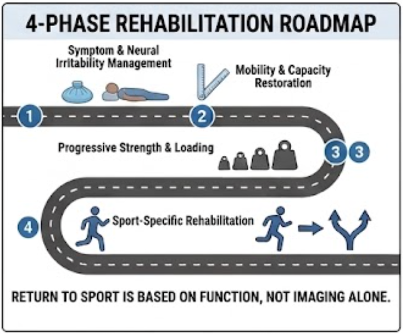
Phase 1: Symptom and Neural Irritability Management
Early rehabilitation should reduce highly aggravating loading while maintaining tolerable activity.
Priorities may include:
- Managing symptom-provoking sport exposure
- Avoiding excessive neurological irritation
- Maintaining appropriate activity
- Gradually restoring tolerated mobility
The goal is not prolonged complete inactivity.
It is to find the amount of movement and loading the athlete can currently tolerate.
Phase 2: Restore Mobility and Physical Capacity
As symptoms stabilize, rehabilitation should address relevant deficits in:
- Lumbar mobility
- Hip mobility
- Trunk function
- Balance
- General movement tolerance
An important consideration is that subjective improvement may occur before objective recovery of strength or spinal mobility.
In athletes, feeling better does not necessarily mean that sport capacity has fully returned.⁸
Phase 3: Progressive Strength and Loading
Rehabilitation can then progress toward increasing:
- Trunk strength
- Trunk endurance
- Hip capacity
- Lower-extremity strength
- Functional lifting and loading tolerance
The objective should not be to identify one universal “core exercise.”
The objective is to progressively rebuild the physical capacity required for the athlete’s sport.
Phase 4: Sport-Specific Rehabilitation
The athlete should eventually progress toward the demands of the actual sport, which may include:
- Running
- Jumping
- Rotational tasks
- Lifting
- Throwing
- Contact preparation
Return-to-sport assessment should integrate:
- Pain
- Neurological symptoms
- Motor and sensory findings
- Mobility
- Strength
- Trunk endurance
- Balance
- Sport-specific loading tolerance
A fixed timeline alone should not determine readiness.
When Is Surgery Considered?
Surgery is generally not the first-line treatment for most uncomplicated cases.
It may become appropriate when there is:
- An urgent neurological indication
- Progressive neurological deficit
- Failure of an adequate course of conservative management
- Persistent function-limiting symptoms that prevent meaningful sport participation³,⁶
Interestingly, a systematic review and meta-analysis of elite athletes reported return-to-play rates of approximately:
- 83.0% after operative treatment
- 81.5% after nonoperative treatment
with no significant overall difference in return-to-play time between groups.⁶
Therefore:
surgery should not automatically be assumed to provide a superior return-to-sport outcome simply because the patient is an athlete.
Treatment selection should depend on the neurological presentation, symptom severity, clinical course, sport demands, and response to conservative care.
< Summary >
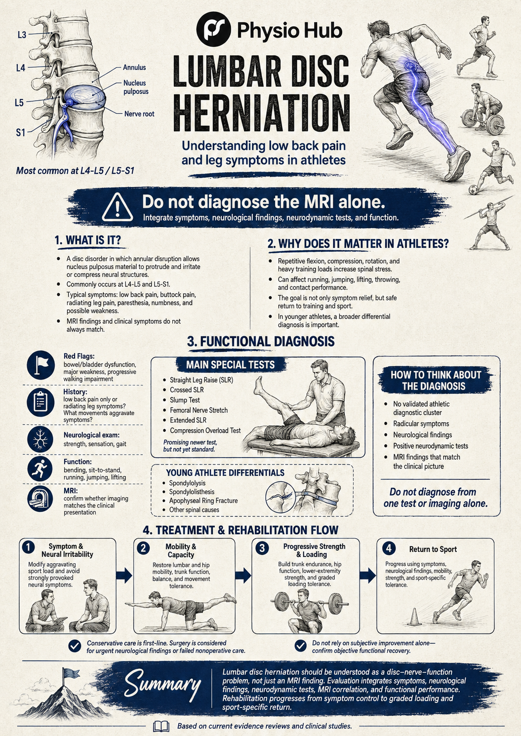
Lumbar disc herniation is more than an abnormal disc seen on MRI.
Clinically, it is best understood as a potential disc–nerve–function problem in which structural findings must be interpreted together with neurological symptoms and athletic function.
Functional diagnosis should progress through:
symptom behavior → neurological examination → neurodynamic testing → functional assessment → MRI correlation.
Rehabilitation generally progresses through:
symptom control → mobility and capacity restoration → progressive strength and loading → sport-specific rehabilitation → return to sport.
For athletes, the final objective is not merely to eliminate back or leg pain.
It is to restore sufficient neurological and physical capacity to tolerate the high spinal and whole-body loads required by sport again.
<PDF file>
Read More



< Reference >
- Du S, Cui Z, Peng S, et al. Clinical efficacy of exercise therapy for lumbar disc herniation: a systematic review and meta-analysis of randomized controlled trials. Front Med (Lausanne). 2025;12:1531637. doi:10.3389/fmed.2025.1531637.
- Hossain MS, Akter S, Siddique MAE, et al. Multidisciplinary conservative treatment outcomes of in-patient physiotherapy set-up among patients with lumbar disc herniation in Dhaka City, Bangladesh: a retrospective, cross-sectional study. J Multidiscip Healthc. 2023;16:587-601. doi:10.2147/JMDH.S400021.
- Hossain MS, Akter S, Siddique MAE, et al. Multidisciplinary conservative treatment outcomes of in-patient physiotherapy set-up among patients with lumbar disc herniation in Dhaka City, Bangladesh: a retrospective, cross-sectional study. J Multidiscip Healthc. 2023;16:587-601. doi:10.2147/JMDH.S400021.
- Al-Sharaa M, Salo SA, Kareem AF, Al-Edanni MS. Slump test versus straight leg raise test in the diagnosing of lumbar disc herniation: a prospective comparative study. Published 2021. doi:10.47723/kcmj.v17i1.309.
- Nee RJ, Coppieters MW, Boyd BS. Reliability of the straight leg raise test for suspected lumbar radicular pain: a systematic review with meta-analysis. Musculoskelet Sci Pract. 2022;59:102529. doi:10.1016/j.msksp.2022.102529.
- Sedrak P, Shahbaz M, Gohal C, Madden K, Aleem I, Khan M. Return to play after symptomatic lumbar disc herniation in elite athletes: a systematic review and meta-analysis of operative versus nonoperative treatment. Sports Health. 2021;13(5):446-453. doi:10.1177/1941738121991782.
- Zhang J, Zhang W, Yue W, Qin W, Li Z, Xu G. Adolescent lumbar disc herniation: etiology, diagnosis, and treatment options. J Orthop Surg Res. 2025;20(1):605. doi:10.1186/s13018-025-06024-3.
- Miñambres-Martín D, Martín-Casas P, López-de-Uralde-Villanueva I, Fernández-de-las-Peñas C, Valera-Calero JA, Plaza-Manzano G. Physical function in amateur athletes with lumbar disc herniation and chronic low back pain: a case-control study. Int J Environ Res Public Health. 2022;19(6):3743. doi:10.3390/ijerph19063743.
- Pesonen J, Shacklock M, Suomalainen JS, et al. Extending the straight leg raise test for improved clinical evaluation of sciatica: validity and diagnostic performance with reference to magnetic resonance imaging. BMC Musculoskelet Disord. 2021;22(1):808. doi:10.1186/s12891-021-04649-z.
- Al-Sharaa M, Salo SA, Kareem AF, Al-Edanni MS. Slump test versus straight leg raise test in the diagnosing of lumbar disc herniation: a prospective comparative study. Published 2021. doi:10.47723/kcmj.v17i1.309.
- Madhesh M, Eapen C, Eappakkam Kumaraswamy P, Palaniswamy V. Diagnostic accuracy of the compression overload test versus straight leg raise test in detecting lumbar disc herniation: an MRI-validated cross-sectional study. Eur Spine J. 2025;34(10):4377-4385. doi:10.1007/s00586-025-09164-6.
Stay Informed with Physio Hub
Unlock a world of knowledge with our expertly curated articles on fitness, rehabilitation, and health & diet. Stay ahead with the latest evidence-based insights and practical advice. Subscribe now to receive updates and never miss out on new content!